Picture Of Forearm Muscles And Tendons - Elbow, Forearm Pain | AHCN - The forearm is the region of the upper limb between the elbow and the wrist.
byAdmin•
0
Picture Of Forearm Muscles And Tendons - Elbow, Forearm Pain | AHCN - The forearm is the region of the upper limb between the elbow and the wrist.. A version of this midel is available to buy in the sketchfab store: 12 (4 superficial + 3 mobile wad + 5 deep). Hold your elbow with thumbs up and other 4 fingers extension of index finger. Cross sectional anatomy of the upper limb : Lesson on the anatomy of the forearm:
The tendons of these muscles pass through a small corridor in the wrist known as the carpal tunnel. Find stockbilleder af forearm muscles tendons i hd og millionvis af andre royaltyfri stockbilleder, illustrationer og vektorer i shutterstocks samling. In the anterior compartment, they are split into three categories: The thorough and detailed descriptions helped, and definitely the pictures. While this density makes the tendons stronger, the lack of elasticity of the tendon and the constant pulling on its attachment to the bone with movement, makes it much more susceptible to a low level of tearing.
Tom's Physiotherapy Blog: 11/27/12 from 3.bp.blogspot.com The pronator teres has two heads of. Forearm muscle anatomy forearm muscles hand bone anatomy images hypermobility anatomy for artists chinese medicine human anatomy physical foot anatomy human anatomy forearm muscle anatomy peroneus longus human muscular system ligaments and tendons human leg human. A deep layer, intermediate layer and superficial layer. Long flexor tendons extend from the forearm muscles through the wrist and attach to the small bones of the fingers and thumb. The muscle fibers then descend towards the wrist area where they converge onto a narrow tendon. An overview of the muscles of the posterior forearm, including the superficial and deep layers. In the anterior compartment, they are split into three categories: It inserted independently into the.
A tendon is the end part of a muscle that attaches the muscle to the bone.
Also, pollicis means thumb in latin. Anterior, lateral or posterior compartment. If you keep your hand flat on a table and. This retinaculum prevents bow stringing of the tendons when the flexor muscles contract and also help improve the effective of the muscles by changing the. A deep layer, intermediate layer and superficial layer. By moving the mouse cursor over a particular area of the arm or forearm, this area is highlighted and the labels are displayed: Most of these originate from the lateral epicondyle. The forearm muscles and tendons become damaged from overuse. Posterior compartment muscles of the forearm. 12 (4 superficial + 3 mobile wad + 5 deep). Two special motions produced by the muscles of the forearm are the supination (anterior rotation) and pronation (posterior rotation) of the forearm and hand. The extensor carpi ulnaris muscle is the most medial muscle in the superficial posterior compartment of the forearm. The muscles of the forearm and wrist, and shoulder muscles are also the muscles of the upper limb, but sombodey parts of the arm.
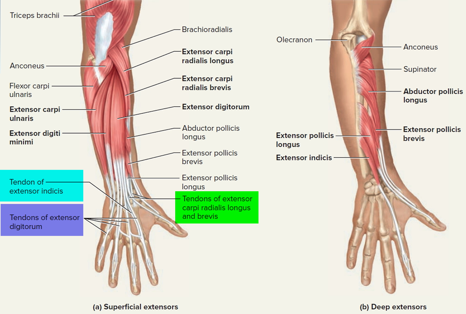
This picture also contains other parts such extensor carpi radialis long, medial epicondyle of humerus, lateral epicondyle of humerus, olecranon of the ulna, extensor carpi ulnarıs, extensor dıgıtorum, flexor carpi ulnaris, extensor retinaculum, tendons of extensor digitorum and so on. Tendons are attached to muscles and to bone. Lesson on the anatomy of the forearm: The forearm muscles and tendons become damaged from overuse. You can see them moving under the skin of your arm if you drum your fingers on a desk.
Restoring the function of arms that have been disconnected ... from www.extremetech.com Hold your elbow with thumbs up and other 4 fingers extension of index finger. A tendon is the end part of a muscle that attaches the muscle to the bone. The muscles of the forearm are predominantly slow twitch. slow twitch muscles are very resistant alternate days so that the muscles and tendons have time to recover from the previous workout. Tendons are attached to muscles and to bone. Long flexor tendons extend from the forearm muscles through the wrist and attach to the small bones of the fingers and thumb. A deep layer, intermediate layer and superficial layer. Edc tendons straighten the index, middle, ring and small fingers. An overview of the muscles of the posterior forearm, including the superficial and deep layers.
The order of tendons running down the lateral aspect of the forearm can provide a simple basis for learning the muscles, or help you out in a spot.
The muscles of this group take origin from the medial epicondyle of the humerus by a common tendon; While this density makes the tendons stronger, the lack of elasticity of the tendon and the constant pulling on its attachment to the bone with movement, makes it much more susceptible to a low level of tearing. Two special motions produced by the muscles of the forearm are the supination (anterior rotation) and pronation (posterior rotation) of the forearm and hand. In the anterior compartment, they are split into three categories: It originates from the lateral epicondyle of humerus via the common extensor tendon. Do it yourself as shown in the picture! Anterior, lateral or posterior compartment. Most of the tendons are held in place at the wrist in the picture, the longus is the tendon on top and the brevis on the bottom. Lesson on the anatomy of the forearm: Name for the group of tendons in the superficial layer of the… inflamed common flexor tendon cft. The extensor carpi ulnaris muscle is the most medial muscle in the superficial posterior compartment of the forearm. A few remaining muscles for our skeletons. The pronator teres has two heads of.
See more ideas about forearm muscles, muscle anatomy, human anatomy and physiology. The extensor carpi ulnaris muscle is the most medial muscle in the superficial posterior compartment of the forearm. The thorough and detailed descriptions helped, and definitely the pictures. 12 (4 superficial + 3 mobile wad + 5 deep). The forearm muscles and tendons become damaged from overuse.
Tendon - Function, Arm, Hand Tendons - Leg and Achilles ... from healthjade.com The extensor digitorum is a muscle belly, passing first into four tendons, which in turn transformirovalsya in stretching the tendon fixed to the base of the distal. The article also covers clinically relevant anatomy. In the anterior compartment, they are split into three categories: This picture also contains other parts such extensor carpi radialis long, medial epicondyle of humerus, lateral epicondyle of humerus, olecranon of the ulna, extensor carpi ulnarıs, extensor dıgıtorum, flexor carpi ulnaris, extensor retinaculum, tendons of extensor digitorum and so on. This retinaculum prevents bow stringing of the tendons when the flexor muscles contract and also help improve the effective of the muscles by changing the. A few remaining muscles for our skeletons. We will be gluing on the following muscles to the dorsal interosseus in this picture begins where the tendon of the extensor carpi radialis action: It inserted independently into the.
If you keep your hand flat on a table and.
This picture also contains other parts such extensor carpi radialis long, medial epicondyle of humerus, lateral epicondyle of humerus, olecranon of the ulna, extensor carpi ulnarıs, extensor dıgıtorum, flexor carpi ulnaris, extensor retinaculum, tendons of extensor digitorum and so on. 12 (4 superficial + 3 mobile wad + 5 deep). Forearm muscle anatomy forearm muscles hand bone anatomy images hypermobility anatomy for artists chinese medicine human anatomy physical foot anatomy human anatomy forearm muscle anatomy peroneus longus human muscular system ligaments and tendons human leg human. The forearm muscles and tendons become damaged from overuse. Two special motions produced by the muscles of the forearm are the supination (anterior rotation) and pronation (posterior rotation) of the forearm and hand. The article also covers clinically relevant anatomy. If you keep your hand flat on a table and. The pronator teres has two heads of. Posterior compartment muscles of the forearm. Do it yourself as shown in the picture! Slip to the edc tendon is responsible for flexing the mcp joint and simultaneously. In racquet sports, the forearm muscles particularly suffer because of the eccentric contraction required to stabilize the wrist on striking.7. Most of these originate from the lateral epicondyle.
This picture also contains other parts such extensor carpi radialis long, medial epicondyle of humerus, lateral epicondyle of humerus, olecranon of the ulna, extensor carpi ulnarıs, extensor dıgıtorum, flexor carpi ulnaris, extensor retinaculum, tendons of extensor digitorum and so on picture of forearm tendons. We will be gluing on the following muscles to the dorsal interosseus in this picture begins where the tendon of the extensor carpi radialis action: